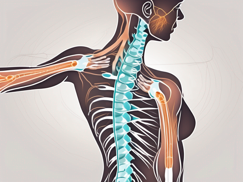
which muscles are innervated by the spinal accessory nerve

The spinal accessory nerve, also known as cranial nerve XI, is a vital component of the human nervous system. It plays a crucial role in innervating specific muscles that contribute to various movements and actions. Understanding the anatomy, function, and disorders related to the spinal accessory nerve can provide valuable insights into the importance of this nerve in our daily lives, as well as in physical therapy. In this article, we will explore the muscles innervated by the spinal accessory nerve and discuss its role in movement and rehabilitation.
Understanding the Spinal Accessory Nerve
The spinal accessory nerve is a crucial component of the nervous system, playing a vital role in the movement and coordination of the neck, shoulders, and head. Let’s delve deeper into the anatomy and function of this remarkable nerve.
Anatomy of the Spinal Accessory Nerve
The spinal accessory nerve, also known as cranial nerve XI, originates from the upper cervical spinal roots in the upper neck region. It is a paired nerve, meaning it exists on both sides of the body, ensuring bilateral control and coordination.
After its origin, the spinal accessory nerve travels upwards, traversing through the intricate network of bones in the skull. It then joins forces with the cranial nerves, forming a complex network of neural pathways.
Within this network, the spinal accessory nerve splits into internal and external branches, each serving distinct functions. The internal branch innervates the muscles responsible for controlling the soft palate and vocal cords, contributing to speech and swallowing. On the other hand, the external branch supplies motor fibers to the trapezius muscle and sternocleidomastoid muscle.
The trapezius muscle, located in the upper back and neck, plays a crucial role in stabilizing and moving the shoulder blades. It allows for movements such as shrugging the shoulders, extending the head, and rotating the neck. The sternocleidomastoid muscle, situated in the front of the neck, facilitates head rotation and flexion.
Function of the Spinal Accessory Nerve
The spinal accessory nerve primarily functions as a motor nerve, controlling the movement of specific muscles involved in various actions. It works in conjunction with other nerves and muscles to provide stability and coordination during movements of the neck, shoulders, and head.
When the spinal accessory nerve is functioning optimally, it allows for smooth and coordinated movements of the neck, shoulders, and head. For example, when you turn your head to look over your shoulder while driving, the spinal accessory nerve is responsible for the precise coordination of the muscles involved in this movement.
In addition to its motor function, the spinal accessory nerve also plays a role in proprioception, which is the body’s ability to sense its position in space. This sensory information is crucial for maintaining balance and posture.
Damage or dysfunction of the spinal accessory nerve can lead to various symptoms, such as weakness or paralysis of the trapezius muscle and sternocleidomastoid muscle. This can result in difficulties with shoulder movements, neck rotation, and head flexion.
Understanding the intricate anatomy and function of the spinal accessory nerve highlights its importance in our daily activities. From simple actions like turning our heads to more complex movements involving the shoulders, this nerve ensures smooth coordination and stability. Appreciating the complexity of our nervous system allows us to better understand and care for our bodies.
Muscles Innervated by the Spinal Accessory Nerve
The Sternocleidomastoid Muscle
One of the main muscles innervated by the spinal accessory nerve is the sternocleidomastoid muscle, or SCM for short. This muscle is located on the sides of the neck and plays a crucial role in head movement and posture. Contraction of the SCM muscle allows for rotation and tilting of the head, while contraction on both sides together enables flexion of the neck.
The sternocleidomastoid muscle originates from two points: the sternum (breastbone) and the clavicle (collarbone). It then inserts onto the mastoid process, which is a bony prominence behind the ear. The muscle is divided into two parts, the sternal head and the clavicular head, which work together to perform their functions.
When the sternocleidomastoid muscle contracts on one side, it causes ipsilateral (same side) rotation of the head. For example, if the left SCM contracts, it will rotate the head to the left. Conversely, contraction of both SCM muscles together results in flexion of the neck, bringing the chin towards the chest. This movement is commonly seen when someone is trying to touch their chin to their chest or during certain exercises that target the neck muscles.
The Trapezius Muscle
The trapezius muscle is another significant muscle innervated by the spinal accessory nerve. It is a large muscle that covers the upper back and neck. The trapezius muscle plays a crucial role in shoulder movement and stability. Contraction of the trapezius muscle allows for elevation, retraction, and depression of the shoulders. It also helps with head movement and maintaining proper posture.
The trapezius muscle has a broad origin, with fibers extending from the occipital bone at the base of the skull, the spinous processes of the cervical and thoracic vertebrae, and the ligamentum nuchae (a fibrous structure in the neck). From its origin, the muscle fans out and inserts onto the clavicle, the acromion process of the scapula (shoulder blade), and the spine of the scapula.
Due to its extensive attachments, the trapezius muscle is involved in various movements of the shoulder girdle. Contraction of the upper fibers of the trapezius muscle results in elevation of the shoulders, such as when shrugging. The middle fibers retract the scapulae, pulling them towards the spine. The lower fibers depress the scapulae, allowing for movements like lowering the shoulders after shrugging or during certain exercises that target the trapezius muscle.
In addition to its role in shoulder movement, the trapezius muscle also assists in head movement. When both trapezius muscles contract together, they extend the neck, pulling the head backward. This movement is commonly observed when someone is trying to look up at the ceiling or during exercises that involve neck extension.
The Role of the Spinal Accessory Nerve in Movement
Movement of the Neck and Shoulders
The spinal accessory nerve, also known as cranial nerve XI, is a crucial component in the movement and coordination of the neck and shoulders. Along with the muscles it innervates, this nerve allows for a wide range of movements, providing both strength and precision to these areas of the body.
Working in harmony with other muscles and nerves, the spinal accessory nerve enables various movements of the neck, including flexion, extension, rotation, and lateral bending. These movements are essential for everyday activities such as looking around, nodding, and tilting the head.
Moreover, the spinal accessory nerve plays a vital role in the movement of the shoulders. It contributes to the elevation, depression, and retraction of the shoulders, allowing for a wide range of arm movements. Whether it’s reaching for an object on a high shelf or pulling something towards you, the spinal accessory nerve is at work, ensuring the smooth coordination of these actions.
Role in Head Rotation and Shrugging
One of the specific functions of the spinal accessory nerve is its significant contribution to the rotation of the head and the shrugging of the shoulders. When we rotate our head from side to side, the coordinated action of both sides of the sternocleidomastoid muscles, which are innervated by the spinal accessory nerve, is crucial. These muscles work in unison to allow for smooth and controlled head rotation, enabling us to look around and engage with our surroundings.
Similarly, when we shrug our shoulders, the activation of the trapezius muscles, which are also innervated by the spinal accessory nerve, is essential. The trapezius muscles play a significant role in shoulder movement and stability, allowing us to lift and lower our shoulders effortlessly. Whether we are expressing uncertainty or simply adjusting our backpack straps, the spinal accessory nerve ensures the proper functioning of these movements.
Understanding the role of the spinal accessory nerve in movement is crucial not only for medical professionals but also for individuals seeking to maintain their overall well-being. By appreciating the intricate workings of this nerve, we can better understand the importance of proper posture, exercise, and rehabilitation techniques that promote the health and functionality of the neck and shoulders.
Disorders Related to the Spinal Accessory Nerve
The spinal accessory nerve, also known as cranial nerve XI, plays a crucial role in controlling certain muscles involved in movement of the head and shoulders. Injury or damage to this nerve can lead to various symptoms and limitations in movement.
Symptoms of Spinal Accessory Nerve Damage
When the spinal accessory nerve is damaged, it can result in weakness or paralysis of the muscles innervated by this nerve. This can make it difficult for individuals to rotate their head, lift their shoulders, or maintain proper posture. Simple tasks like turning your head to check for oncoming traffic or lifting objects above your head can become challenging.
In more severe cases, individuals may experience muscle atrophy, which is the wasting away or loss of muscle tissue. This can further contribute to weakness and limited range of motion in the affected areas. Additionally, pain may be present in the neck, shoulder, or upper back region.
Treatment and Recovery Options
If you suspect spinal accessory nerve damage or experience any of the mentioned symptoms, it is crucial to seek medical advice promptly. A healthcare professional, such as a neurologist or physical therapist, can perform a thorough evaluation and provide guidance on appropriate treatment options.
Treatment for spinal accessory nerve damage may involve a combination of strengthening exercises, stretching, and physical therapy modalities. These interventions aim to improve muscle strength, increase range of motion, and enhance overall function. Physical therapists can design a personalized rehabilitation plan tailored to the individual’s specific needs and goals.
In rare cases where conservative treatments are not effective, surgical intervention may be considered. This can involve repairing or reconstructing the damaged nerve to restore its function. However, surgery is typically reserved for severe cases or when other treatment options have been exhausted.
Recovery from spinal accessory nerve damage can vary depending on the extent of the injury and individual factors. It is important to follow the recommended treatment plan and engage in regular physical therapy sessions to optimize the chances of recovery. With proper care and rehabilitation, many individuals are able to regain function and improve their quality of life.
The Importance of the Spinal Accessory Nerve in Physical Therapy
The spinal accessory nerve, also known as cranial nerve XI, is a crucial component of the peripheral nervous system. It originates from the upper spinal cord and innervates various muscles in the neck and shoulder region. This nerve plays a significant role in movement, posture, and overall function.
Rehabilitation Exercises for Spinal Accessory Nerve Damage
In physical therapy, the importance of the spinal accessory nerve becomes evident in the rehabilitation of individuals with nerve-related injuries or conditions. When this nerve is damaged, it can lead to weakness, limited range of motion, and difficulties in performing daily activities.
Physical therapists are skilled in designing tailored exercise programs that target the muscles innervated by the spinal accessory nerve. These exercises aim to promote strength, coordination, and mobility. By focusing on specific muscle groups, physical therapy can help individuals regain optimal movement patterns and functionality.
Rehabilitation exercises for spinal accessory nerve damage may include a combination of stretching, strengthening, and coordination exercises. Stretching exercises help improve flexibility and increase the range of motion in the affected muscles. Strengthening exercises, on the other hand, focus on building muscle strength to compensate for any weakness caused by the nerve damage. Coordination exercises aim to improve the synchronization of muscle movements, allowing for smoother and more efficient motor control.
Additionally, physical therapists may incorporate other therapeutic interventions, such as manual therapy techniques, electrical stimulation, or ultrasound, to enhance the effectiveness of the rehabilitation program. These interventions can help reduce pain, inflammation, and muscle spasms, facilitating the healing process.
Preventing Spinal Accessory Nerve Injuries
While accidents or conditions beyond our control can lead to spinal accessory nerve injuries, certain precautions can help reduce the risk. Maintaining proper posture is essential, as poor posture can place excessive strain on the neck and shoulder muscles, potentially damaging the nerve over time.
Practicing safe lifting techniques is another crucial aspect of preventing nerve injuries. When lifting heavy objects, it is important to engage the appropriate muscles and avoid putting excessive stress on the neck and shoulders. Using proper body mechanics and seeking assistance when needed can significantly reduce the risk of nerve damage.
Furthermore, being mindful of excessive strain on the neck and shoulders during daily activities or sports is essential. It is important to listen to your body and take breaks when necessary. If you participate in physical activities that involve the neck or shoulder muscles, such as weightlifting or contact sports, it is crucial to properly warm up and stretch before engaging in these activities. This helps prepare the muscles for the demands of the activity and reduces the risk of injury.
If you have any concerns or questions regarding your specific situation, it is always advisable to consult with a healthcare professional or physical therapist. They can provide personalized guidance and recommendations based on your individual needs and circumstances.
In conclusion, the muscles innervated by the spinal accessory nerve play a vital role in movement, posture, and overall function. Understanding the anatomy, function, and potential disorders related to this nerve can provide valuable insights into its importance in our daily lives. If you experience any symptoms of spinal accessory nerve damage, such as weakness, limited range of motion, or difficulties in performing daily activities, it is crucial to consult with a healthcare professional for accurate diagnosis and appropriate treatment options.
Physical therapy can be an effective tool in rehabilitating spinal accessory nerve injuries, helping individuals regain strength, coordination, and optimal movement patterns. By taking preventive measures, such as maintaining proper posture, practicing safe lifting techniques, and being mindful of excessive strain on the neck and shoulders, we can prioritize the health and well-being of our spinal accessory nerve and the muscles it innervates.


