what part of cervical plexus is the psinal accessory nerve from

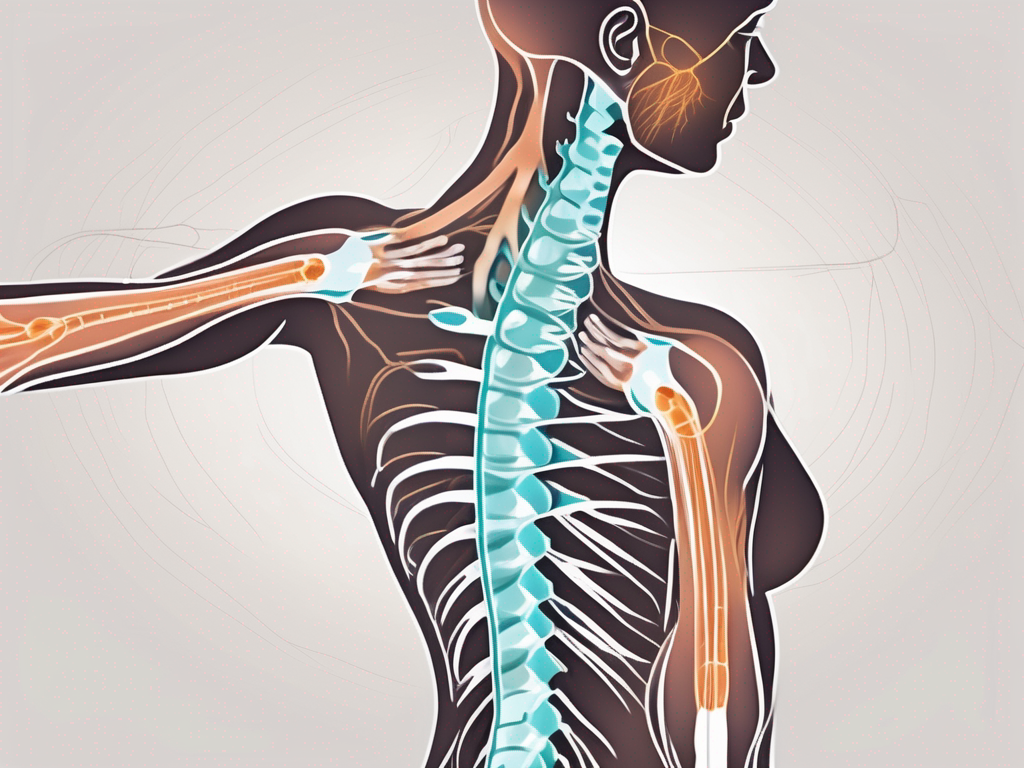
The cervical plexus is a complex network of nerves located in the neck region, specifically in the area between the first cervical vertebra (C1) and the fourth cervical vertebra (C4). This intricate structure consists of several branches that innervate different areas of the neck, shoulder, and upper chest. Among these branches is the spinal accessory nerve, which plays an essential role in motor function and sensation.
Understanding the Cervical Plexus
The cervical plexus is a complex network of nerves that plays a crucial role in the functioning of various muscles and sensory organs in the neck and upper chest region. It is formed by the merging of nerve fibers from the ventral rami of the first four cervical spinal nerves: C1 to C4. This intricate web of nerves is responsible for transmitting important signals and information throughout the neck and upper body.
Within the cervical plexus, there are multiple branches, each with a specific function and target area. These branches include the lesser occipital nerve, great auricular nerve, transverse cervical nerve, supraclavicular nerves, and the phrenic nerve. Each branch serves a unique purpose, innervating different regions of the neck and providing both motor and sensory information.
Anatomy of the Cervical Plexus
The lesser occipital nerve, one of the branches of the cervical plexus, is responsible for providing sensory information to the skin of the scalp and behind the ear. It allows us to perceive sensations such as touch, temperature, and pain in these areas. The great auricular nerve, another branch, supplies sensory information to the skin of the ear and the area just below it. This nerve enables us to feel sensations such as pressure and temperature in these regions.
The transverse cervical nerve, as its name suggests, runs horizontally across the neck. It provides sensory information to the skin on the anterior and lateral aspects of the neck. This nerve allows us to feel sensations such as touch, pressure, and temperature in these areas. The supraclavicular nerves, on the other hand, supply sensory information to the skin over the clavicle and the upper chest region. These nerves play a role in our ability to perceive touch, pressure, and temperature in these specific areas.
Perhaps one of the most significant branches of the cervical plexus is the phrenic nerve. This nerve originates from the C3 to C5 spinal nerves and provides motor control to the diaphragm, the primary muscle responsible for breathing. Without the phrenic nerve, the diaphragm would not be able to contract and relax properly, leading to difficulties in breathing.
Functions of the Cervical Plexus
The cervical plexus serves several important functions in the neck and upper body. One of its primary functions is to provide motor control to the muscles of the neck and upper shoulder region. Through its various branches, the cervical plexus enables movements such as turning the head from side to side and shrugging the shoulders. These movements are essential for our everyday activities and range of motion.
In addition to motor control, the cervical plexus also plays a crucial role in sensory perception. It provides tactile sensations to the skin of the neck, allowing us to feel touch, pressure, and temperature in this area. Furthermore, the cervical plexus transmits sensory information from the organs in the head and neck, allowing us to perceive sensations such as pain, temperature, and pressure from these regions. This sensory feedback is vital for our overall well-being and awareness of our surroundings.
In conclusion, the cervical plexus is a complex network of nerves that plays a vital role in the functioning of various muscles and sensory organs in the neck and upper chest region. Its branches provide both motor control and sensory information, allowing us to perform essential movements and perceive sensations in these areas. Understanding the anatomy and functions of the cervical plexus helps us appreciate the intricate mechanisms that enable us to move and experience the world around us.
The Spinal Accessory Nerve Explained
The spinal accessory nerve, also known as cranial nerve XI, is one of the branches of the cervical plexus. Unlike the other branches, which primarily provide sensation and motor control to the neck and head, the spinal accessory nerve has a more specific function related to the movement of certain muscles in the shoulder and neck.
The spinal accessory nerve plays a crucial role in the coordination of movements involving the head, neck, and shoulder. It is responsible for transmitting signals from the brain to the sternocleidomastoid and trapezius muscles, enabling precise control and coordination of these muscles.
Anatomy of the Spinal Accessory Nerve
The spinal accessory nerve originates from the upper spinal cord, specifically from the motor neurons located in the ventral horn of the spinal cord segments C1 to C5. These motor neurons are responsible for initiating and controlling voluntary movements.
From its origin, the spinal accessory nerve travels upward, passing through the foramen magnum, which is a large opening at the base of the skull. Inside the skull, the spinal accessory nerve joins forces with another cranial nerve, the vagus nerve, forming a complex network of nerves.
After joining the vagus nerve, the spinal accessory nerve emerges through the jugular foramen, a narrow opening located at the base of the skull. This exit point allows the nerve to continue its journey downward, extending towards the neck and shoulder region.
Functions of the Spinal Accessory Nerve
Once the spinal accessory nerve exits the skull through the jugular foramen, it continues its journey downward, where it innervates specific muscles in the neck and shoulder. The primary muscles controlled by this nerve are the sternocleidomastoid muscle and the trapezius muscle.
The sternocleidomastoid muscle, located on both sides of the neck, plays a vital role in head movements. It allows for the rotation and tilting of the head, enabling us to turn our heads from side to side and nod up and down. The spinal accessory nerve provides the necessary signals for the contraction and relaxation of this muscle, allowing for precise control of head movements.
The trapezius muscle, a large muscle that spans the upper back and neck, is responsible for various movements involving the shoulder and scapula. It helps stabilize the shoulder joint, retract the scapula, and elevate or depress the shoulder. The spinal accessory nerve ensures the proper functioning of the trapezius muscle by transmitting signals that initiate and regulate its contractions.
In addition to its role in muscle control, the spinal accessory nerve also plays a part in transmitting sensory information. It carries proprioceptive information from the muscles it innervates, providing feedback to the brain about the position and movement of the head, neck, and shoulder.
Overall, the spinal accessory nerve is a vital component of the nervous system, facilitating precise control and coordination of movements involving the head, neck, and shoulder. Its intricate anatomy and functions highlight the complexity of the human body and the remarkable interplay between nerves and muscles.
The Relationship Between the Cervical Plexus and Spinal Accessory Nerve
Now that we have a basic understanding of the cervical plexus and spinal accessory nerve individually, let’s explore how they are interconnected and their collective role in the body’s functioning.
The cervical plexus, a network of nerves located in the neck, plays a crucial role in innervating various muscles and structures in the head, neck, and upper shoulders. It consists of nerve fibers originating from the upper cervical segments of the spinal cord, primarily from C1 to C5. One of the major branches that emerges from this plexus is the spinal accessory nerve.
How the Spinal Accessory Nerve Originates from the Cervical Plexus
The spinal accessory nerve, also known as cranial nerve XI, arises from the nerve fibers of the upper cervical segments of the spinal cord. These nerve fibers merge together within the cervical plexus, forming a complex network of connections. As they converge, they give rise to the spinal accessory nerve, which then exits the skull through the jugular foramen.
Once it exits the skull, the spinal accessory nerve takes a unique path, traveling down the neck and branching out to innervate specific muscles in the shoulder and neck region. This intricate pathway highlights the intimate relationship between the spinal accessory nerve and the cervical plexus.
The Role of the Spinal Accessory Nerve in the Cervical Plexus
While the spinal accessory nerve is technically a branch of the cervical plexus, it serves as an important connection between the cervical plexus and the muscles of the shoulder and neck. Its primary function is to provide motor control to the sternocleidomastoid and trapezius muscles, allowing for precise movements and maintaining proper posture.
The sternocleidomastoid muscle, located in the front of the neck, is responsible for various movements of the head and neck, including rotation and flexion. The trapezius muscle, on the other hand, spans across the upper back and neck, playing a crucial role in shoulder movement and stability.
Without the spinal accessory nerve’s innervation, these muscles would not be able to function properly, leading to difficulties in head and neck movements, as well as compromised shoulder stability. The intricate interplay between the spinal accessory nerve and the cervical plexus ensures the coordination and precision of these movements.
In addition to its motor function, the spinal accessory nerve also carries sensory information from the muscles it innervates, providing feedback to the central nervous system about the position and tension of these muscles. This feedback loop allows for fine-tuning of movements and helps maintain balance and coordination.
In conclusion, the relationship between the cervical plexus and spinal accessory nerve is a fascinating example of the intricate connections within the human body. The cervical plexus serves as the origin of the spinal accessory nerve, while the spinal accessory nerve plays a crucial role in providing motor control and sensory feedback to the muscles of the shoulder and neck. Understanding this relationship enhances our knowledge of the complex functioning of the human body.
Medical Significance of the Spinal Accessory Nerve and Cervical Plexus
The spinal accessory nerve and cervical plexus hold significant medical importance, as disorders affecting these structures can cause complications and affect daily life. It is essential to be aware of potential conditions and seek appropriate medical attention for proper diagnosis and treatment.
The spinal accessory nerve, also known as cranial nerve XI, plays a crucial role in the innervation of certain muscles in the neck and shoulder region. It originates from the upper spinal cord and travels through the neck, providing motor function to the trapezius and sternocleidomastoid muscles. These muscles are responsible for various movements, such as rotating and tilting the head, as well as elevating and retracting the shoulders.
Disorders affecting the spinal accessory nerve can have a significant impact on a person’s ability to perform daily activities. Nerve compression or injury, which can occur due to trauma or repetitive motion, is a common cause of spinal accessory nerve dysfunction. This can result in weakness, pain, and limited range of motion in the neck and shoulder area.
Diagnosing disorders involving the spinal accessory nerve often requires a comprehensive evaluation by a healthcare professional. They may perform a physical examination, assess the patient’s medical history, and order additional tests, such as electromyography (EMG) or nerve conduction studies, to determine the extent of nerve damage.
Conditions Affecting the Spinal Accessory Nerve
Some common conditions that can affect the function of the spinal accessory nerve include nerve compression or injury, which can occur due to trauma or repetitive motion. Symptoms may include weakness, pain, and limited range of motion in the neck and shoulder area. If you suspect any issues with your spinal accessory nerve, it is crucial to consult with a healthcare professional for a proper diagnosis and guidance.
One condition that can affect the spinal accessory nerve is called accessory nerve palsy. This condition occurs when the nerve is damaged or compressed, leading to weakness or paralysis of the trapezius and sternocleidomastoid muscles. Accessory nerve palsy can result from various causes, including trauma, surgical procedures, or tumors in the neck region.
Another condition that can affect the spinal accessory nerve is thoracic outlet syndrome. This syndrome occurs when the nerves and blood vessels in the neck and shoulder area are compressed or irritated. It can cause pain, numbness, and tingling in the upper extremities, as well as weakness and muscle wasting in the affected arm.
Treatment and Management of Disorders Involving the Cervical Plexus and Spinal Accessory Nerve
Treatment options for conditions affecting the spinal accessory nerve or cervical plexus vary depending on the specific diagnosis and severity of the condition. It may involve physical therapy, medication, or, in some cases, surgical intervention. Seeking professional medical advice from a qualified healthcare provider is crucial to ensure appropriate treatment and management.
Physical therapy plays a vital role in the rehabilitation of individuals with spinal accessory nerve disorders. Therapists can design specific exercises and stretches to improve muscle strength, range of motion, and functional abilities. They may also incorporate modalities such as heat or cold therapy, electrical stimulation, or manual techniques to alleviate pain and promote healing.
In some cases, medications may be prescribed to manage pain and inflammation associated with spinal accessory nerve disorders. Nonsteroidal anti-inflammatory drugs (NSAIDs), muscle relaxants, and analgesics can provide temporary relief and improve overall comfort. However, it is essential to follow the healthcare provider’s instructions and monitor for any potential side effects.
Surgical intervention may be necessary in severe cases of spinal accessory nerve disorders that do not respond to conservative treatments. The specific surgical procedure will depend on the underlying cause and may involve decompression of the nerve, repair of damaged structures, or nerve grafting.
It is important to note that early intervention and proper management can significantly improve the prognosis for individuals with spinal accessory nerve disorders. Therefore, if you experience any symptoms or suspect any issues with your spinal accessory nerve or cervical plexus, it is crucial to seek medical attention promptly for a thorough evaluation and appropriate treatment.
Conclusion: The Importance of Understanding the Cervical Plexus and Spinal Accessory Nerve
In conclusion, the cervical plexus and spinal accessory nerve play integral roles in the functioning of the neck, shoulder, and upper chest. Understanding the anatomy and functions of these structures provides valuable insight into the complexities of the human body. If you have any concerns or experience symptoms related to the cervical plexus or spinal accessory nerve, do not hesitate to consult with a healthcare professional for proper evaluation and guidance tailored to your specific needs.


