how to find the spinal accessory nerve during dissection

The spinal accessory nerve, also known as cranial nerve XI, plays a crucial role in the movement of the neck and shoulders. It is essential for proper functioning and coordination of these muscles. Locating this nerve during dissection can be challenging but is a fundamental skill for anatomists, surgeons, and medical professionals. In this article, we will guide you through the process of finding the spinal accessory nerve during dissection, providing you with valuable insights and tips along the way.
Understanding the Anatomy of the Spinal Accessory Nerve
The spinal accessory nerve, also known as cranial nerve XI, is a vital component of the nervous system. It plays a crucial role in the movement and stability of the neck and shoulder girdle. To fully comprehend its significance, it is essential to explore the intricate details of its origin, pathway, function, and importance.
The Origin and Pathway of the Spinal Accessory Nerve
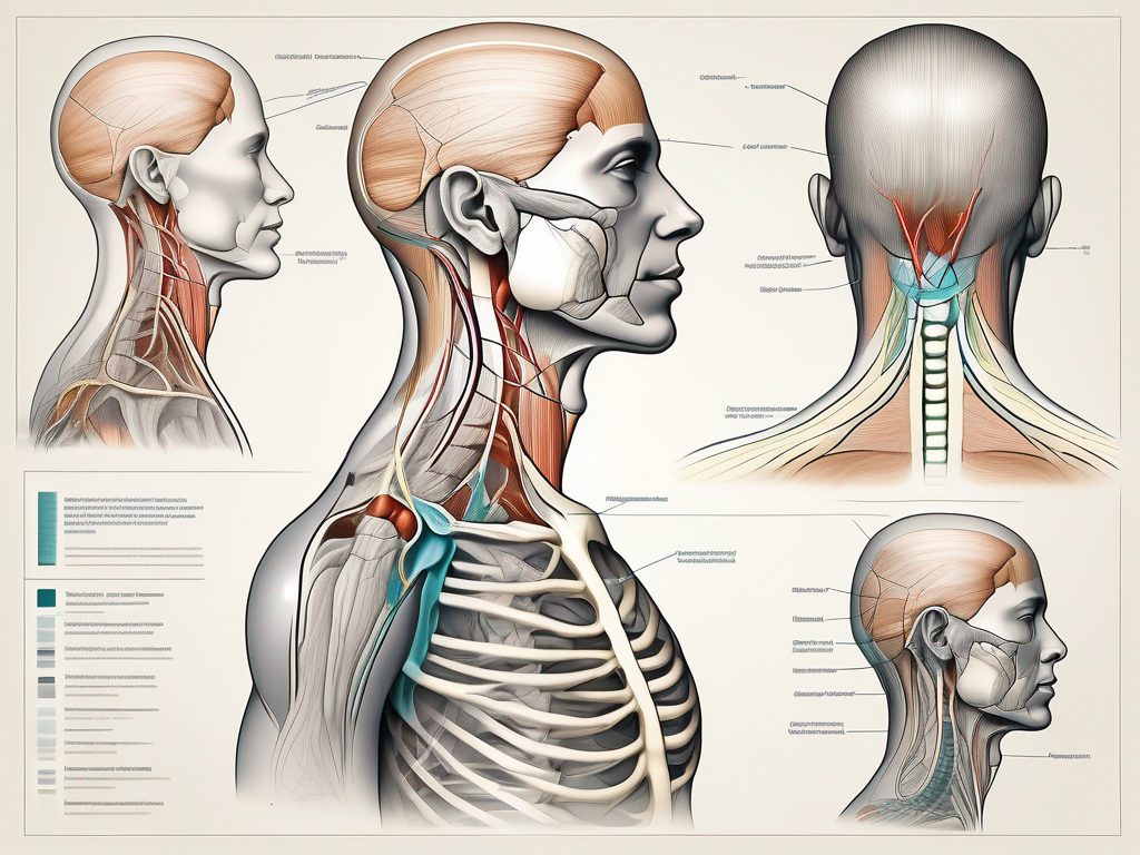
The spinal accessory nerve originates from the upper segments of the spinal cord, specifically the cervical spinal cord. It emerges from the spinal cord at the level of the upper cervical vertebrae. These nerve roots then travel superiorly, passing through the foramen magnum, a large opening at the base of the skull.
Once inside the skull, the spinal accessory nerve exits through the jugular foramen, a narrow passageway located between the temporal and occipital bones. This foramen serves as a gateway for the nerve to leave the protective confines of the skull and venture into the neck region.
As it descends along the posterior triangle of the neck, the spinal accessory nerve takes a deep course beneath the sternocleidomastoid muscle, one of the major muscles responsible for the movement and stabilization of the neck. This deep pathway ensures the nerve’s protection and allows it to reach its destination without interference.
Upon reaching its destination, the spinal accessory nerve branches out and innervates two crucial muscles: the trapezius and sternocleidomastoid muscles. These muscles play a pivotal role in various movements of the neck and shoulder girdle, including rotation, lateral flexion, elevation, retraction, and depression.
The Function and Importance of the Spinal Accessory Nerve
The spinal accessory nerve is responsible for coordinating the movement and stability of the neck and shoulder girdle. Its primary function is to control the rotation and lateral flexion of the neck, allowing for smooth and precise movements.
In addition to neck movements, the spinal accessory nerve also plays a crucial role in the elevation, retraction, and depression of the shoulder blades. These movements are essential for maintaining proper posture, performing various upper body exercises, and executing complex tasks that involve the upper extremities.
Understanding the function and importance of the spinal accessory nerve is vital in the fields of anatomy, physiology, and clinical practice. It allows healthcare professionals, such as surgeons, physiotherapists, and occupational therapists, to accurately identify and assess any potential issues or injuries related to this nerve.
Moreover, a comprehensive understanding of the spinal accessory nerve’s anatomy and function aids in the diagnosis and treatment of conditions affecting the neck and shoulder girdle. By recognizing the nerve’s role in movement and stability, healthcare providers can develop targeted interventions and rehabilitation strategies to optimize patient outcomes.
In conclusion, the spinal accessory nerve is a remarkable structure that contributes significantly to the intricate network of the nervous system. Its origin, pathway, function, and importance are all interconnected, forming a comprehensive understanding of this vital nerve. By delving into the details of its anatomy, we gain valuable insights into the complexity and significance of the spinal accessory nerve.
Preparing for the Dissection
Before starting the dissection, it is necessary to gather the appropriate tools and equipment while taking the necessary safety measures. Proper preparation ensures a smooth and efficient dissection process, allowing for accurate observations and analysis.
When embarking on a spinal accessory nerve dissection, it is crucial to have the right tools and equipment at your disposal. These tools enable you to navigate through the intricate structures of the nerve with precision and care.
Necessary Tools and Equipment for Dissection
When preparing for a spinal accessory nerve dissection, ensure you have the following tools and equipment:
- Scalpel: A sharp surgical knife used for making precise incisions and cuts.
- Dissection scissors: Specialized scissors designed for cutting and separating tissues during dissection.
- Forceps: Surgical tweezers used for gripping and manipulating delicate structures.
- Probes: Thin, pointed instruments used for exploring and dissecting tissues without causing damage.
- Dissection tray: A flat surface or container where the dissection takes place, providing a clean and organized workspace.
Having these tools readily available ensures that you can perform the dissection smoothly and efficiently, without any unnecessary interruptions or delays.
Safety Measures and Precautions
Dissection can be a delicate and potentially hazardous procedure. It is essential to follow safety measures and precautions to avoid accidental injury or contamination. By prioritizing safety, you protect yourself and maintain the integrity of the specimen being dissected.
First and foremost, it is crucial to wear personal protective equipment (PPE) throughout the dissection process. This includes gloves, goggles, and a lab coat. Gloves protect your hands from potential exposure to harmful substances, while goggles shield your eyes from any splashes or flying debris. The lab coat acts as a barrier, preventing any contaminants from coming into contact with your clothing.
Furthermore, maintaining a clean and organized workspace is paramount. A clutter-free environment minimizes the risk of accidents and allows for better focus and concentration. Ensure that all tools and equipment are properly sterilized and arranged in a systematic manner, reducing the chances of cross-contamination.
Additionally, it is essential to handle all specimens and equipment with care and respect. Treat the subject of the dissection as a valuable learning opportunity and approach the task with professionalism and diligence.
By adhering to these safety measures and precautions, you create a conducive environment for a successful and informative dissection experience. Remember, the primary goal of any dissection is to gain a deeper understanding of the subject matter and contribute to scientific knowledge.
Step-by-Step Guide to Locating the Spinal Accessory Nerve
Now that we are prepared, let’s dive into the step-by-step process of locating the spinal accessory nerve during dissection.
Initial Steps and Identifying Key Landmarks
Begin by identifying key landmarks on the neck, such as the sternocleidomastoid muscle and the clavicle. The sternocleidomastoid muscle serves as a valuable landmark, as it covers the spinal accessory nerve.
The sternocleidomastoid muscle, also known as SCM, is a large muscle located on each side of the neck. It originates from the sternum and clavicle and inserts on the mastoid process of the temporal bone. Its primary function is to rotate and flex the head. By palpating the SCM, you can easily locate its borders and determine its position in relation to the spinal accessory nerve.
Once you have identified the SCM, move your attention to the clavicle. The clavicle, or collarbone, is a long bone that connects the sternum to the scapula. It serves as an important landmark for locating the spinal accessory nerve, as it lies just below it.
Now that you have identified these key landmarks, you can proceed with the next steps in locating the spinal accessory nerve.
Techniques for Isolating the Spinal Accessory Nerve
Once the key landmarks are identified, carefully dissect the surrounding tissues to expose the spinal accessory nerve. Use blunt dissection techniques and gentle traction to separate the muscle fibers and identify the nerve, creating sufficient space for visualization.
Blunt dissection involves using a blunt instrument, such as a pair of forceps or a blunt-ended probe, to gently separate the tissues without cutting them. This technique allows for a more controlled dissection, minimizing the risk of damaging the nerve or surrounding structures.
As you perform the blunt dissection, you may encounter other structures in the vicinity of the spinal accessory nerve. These structures can include blood vessels, lymph nodes, and other nerves. Take your time to carefully identify and differentiate these structures from the spinal accessory nerve to avoid any potential complications.
Once you have successfully isolated the spinal accessory nerve, you can proceed with further examination or any necessary procedures. Remember to handle the nerve with care and avoid excessive tension or trauma, as it is a delicate structure responsible for innervating important muscles in the neck and shoulder region.
Common Challenges and How to Overcome Them
During the dissection of the spinal accessory nerve, you may encounter various challenges. However, with proper technique and patience, these challenges can be overcome.
One common challenge that may arise during the dissection of the spinal accessory nerve is the presence of surrounding structures that can obstruct the view. These structures, such as blood vessels or connective tissue, can make it difficult to identify and isolate the nerve. In such cases, it is important to take breaks when needed to maintain focus and prevent fatigue. By stepping away from the dissection table and allowing yourself a moment to rest and regroup, you can approach the challenge with a fresh perspective.
Another challenge that may be encountered is poor lighting or inadequate magnification, which can hinder visualization of the nerve. To overcome this challenge, it is essential to ensure that the dissection area is well-lit and that you have access to appropriate magnification tools. Adequate lighting and magnification not only enhance visibility but also allow for better precision and accuracy during the dissection process.
If you find yourself facing difficulties during the dissection, it can be beneficial to consult with a knowledgeable instructor or an experienced colleague. Their guidance and assistance can provide valuable insights and suggestions for overcoming specific challenges. They may have encountered similar obstacles in the past and can offer alternative techniques or approaches that can help you navigate through the dissection process more effectively.
Troubleshooting Tips for Difficult Dissections
If you encounter difficulties during dissection, consider these troubleshooting tips:
- Take breaks when needed to maintain focus and prevent fatigue.
- Ensure adequate lighting and magnification for better visualization.
- Consult with a knowledgeable instructor or experienced colleague for guidance and assistance.
Remember, dissection is a skill that requires practice and patience. Each challenge you encounter provides an opportunity for growth and improvement. By approaching these challenges with a positive mindset and utilizing the troubleshooting tips mentioned above, you can overcome obstacles and successfully dissect the spinal accessory nerve.
Understanding Variations in Anatomy
Keep in mind that anatomical variations exist among individuals. The location and pathway of the spinal accessory nerve may vary slightly from person to person. Familiarize yourself with these variations through literature or consultation with an experienced anatomist.
By understanding the potential variations in the anatomy of the spinal accessory nerve, you can better prepare yourself for the dissection process. It is important to study the relevant literature and consult with experienced anatomists to gain a comprehensive understanding of the possible variations that may be encountered. This knowledge will enable you to adapt your dissection technique accordingly and navigate through any unexpected anatomical differences that may arise.
Furthermore, being aware of these anatomical variations can also help you appreciate the complexity and uniqueness of each individual’s anatomy. It serves as a reminder that while there may be commonalities in the structure of the spinal accessory nerve, there is also room for individual variation. This understanding can foster a deeper appreciation for the intricacies of the human body and the importance of personalized approaches in medical practice.
Post-Dissection Procedures
Once you have successfully located the spinal accessory nerve, it is essential to follow specific post-dissection procedures to ensure proper handling and preservation.
After the dissection, it is crucial to take additional steps to ensure the integrity and longevity of the spinal accessory nerve. These post-dissection procedures involve proper handling, preservation, and cleaning of tools and workspace.
Proper Handling and Preservation of the Spinal Accessory Nerve
Handle the spinal accessory nerve delicately and with care to avoid damage. The nerve is a delicate structure that requires gentle handling to prevent any potential harm. By using fine-tipped forceps or microsurgical instruments, you can minimize the risk of unintended trauma.
If preservation is necessary, consider using appropriate fixatives or cryopreservation techniques. Fixatives such as formalin can help stabilize the nerve tissue and prevent degradation. Cryopreservation, on the other hand, involves freezing the nerve at ultra-low temperatures to maintain its structural integrity for future studies or analysis.
Furthermore, maintaining proper documentation and labeling is essential to ensure accurate identification later on. By carefully documenting the location and condition of the nerve, you can facilitate further research or educational purposes.
Cleaning and Sterilization of Tools and Workspace
After the dissection, clean and sterilize all tools and the workspace thoroughly. Proper cleaning and sterilization are vital to prevent contamination and ensure a safe working environment for future dissections.
Start by removing any residual tissue or debris from the tools used during the dissection. This can be done by rinsing them under running water or using a mild detergent solution. Pay close attention to hard-to-reach areas, such as the tips of forceps or the crevices of scissors, to ensure complete cleanliness.
Once the tools are free from visible debris, proceed with sterilization. Autoclaving, a process that uses high-pressure steam to kill microorganisms, is commonly employed for sterilizing surgical instruments. Alternatively, chemical sterilization using ethylene oxide gas or hydrogen peroxide vapor can also be effective.
It is equally important to clean and disinfect the workspace where the dissection took place. Wipe down all surfaces with a suitable disinfectant, paying special attention to areas that may have come into direct contact with the nerve or bodily fluids.
By following proper disinfection protocols, you can prevent cross-contamination and maintain a safe environment for future dissections or experiments.
Conclusion: Mastering the Skill of Dissection
Mastering the skill of locating the spinal accessory nerve during dissection requires practice, patience, and ongoing learning.
The Importance of Practice and Continued Learning
Regular practice enhances your familiarity with the anatomical structures and improves your dissection techniques. Engage in workshops, courses, and seminars to stay up-to-date with the latest advancements in anatomical dissection.
Future Applications of Your Dissection Skills
Acquiring proficiency in locating the spinal accessory nerve will not only benefit your current anatomical knowledge but also open doors to various medical and surgical specialties. Harnessing this skill can contribute to your overall expertise and future professional opportunities.
Remember, this article serves as a general guide and does not replace professional medical advice. If you are considering performing a dissection or have any medical concerns, consult with a qualified healthcare professional for proper guidance and supervision.


