the accessory nerve innervates which organ

The accessory nerve, also known as the eleventh cranial nerve or CN XI, plays a crucial role in the innervation of certain muscles. While it doesn’t innervate a specific organ, it provides motor function to several muscles involved in head and neck movements. Understanding the anatomy, functions, clinical significance, and treatment options related to the accessory nerve is important for medical professionals and patients alike.
Understanding the Accessory Nerve
The accessory nerve, also known as cranial nerve XI, plays a crucial role in controlling certain muscles in the body. Let’s delve deeper into the anatomy and functions of this important nerve.
Anatomy of the Accessory Nerve
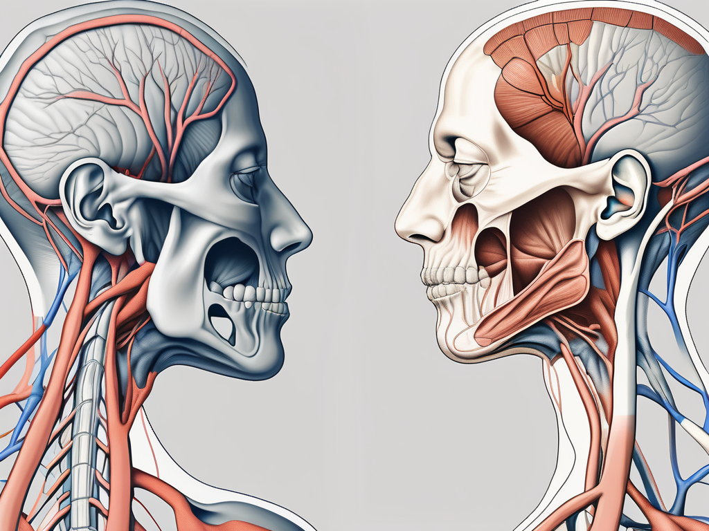
The accessory nerve originates from the upper spinal cord, specifically from the medulla oblongata and the upper cervical spinal cord segments. It emerges as a combination of motor roots that exit the skull through the jugular foramen along with the glossopharyngeal and vagus nerves.
Once the accessory nerve exits the skull, it descends along the posterior aspect of the neck, passing deep to the sternocleidomastoid muscle and giving off branches to innervate different muscles. This intricate network of nerve fibers allows for precise control and coordination of movement.
The path of the accessory nerve through the neck is fascinating. It weaves its way through layers of muscles, blood vessels, and connective tissues, ensuring that it reaches its target muscles with utmost efficiency.
Functions of the Accessory Nerve
The accessory nerve primarily functions to control two important muscles: the sternocleidomastoid and the trapezius.
- Innervation of the Sternocleidomastoid Muscle: The sternocleidomastoid muscle, located on each side of the neck, is responsible for various movements. These include rotation, lateral flexion, and forward flexion of the neck. The accessory nerve provides the necessary motor innervation to ensure the smooth functioning of this muscle.
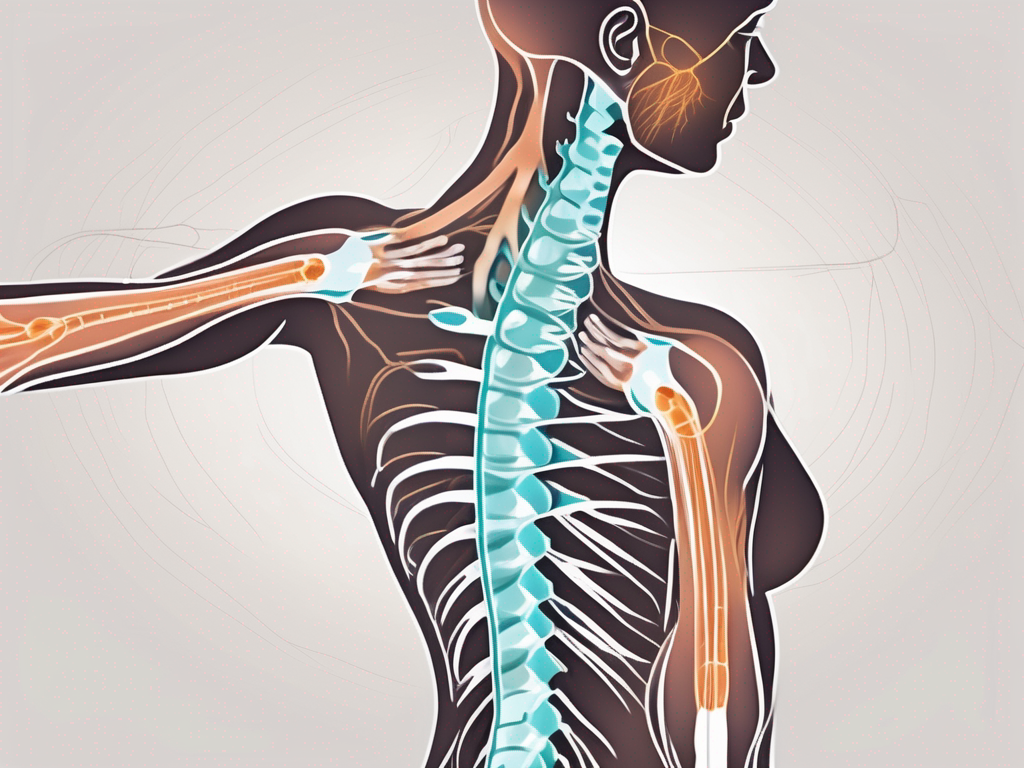
- Innervation of the Trapezius Muscle: The trapezius muscle, which spans the upper back and neck, is another muscle controlled by the accessory nerve. This muscle plays a crucial role in shoulder elevation, retraction, and rotation. Without the innervation from the accessory nerve, these movements would be compromised.
These muscles are vital for proper posture, head movements, and upper limb function. The intricate interplay between the accessory nerve and these muscles allows us to perform a wide range of activities, from turning our heads to lifting objects.
Understanding the accessory nerve and its functions provides us with valuable insights into the complexity of the human body. The precise coordination required for even the simplest movements is truly remarkable.
The Accessory Nerve and the Muscular System
The accessory nerve, also known as cranial nerve XI, plays a vital role in the innervation of various muscles in the neck and shoulder region. This nerve is responsible for supplying motor fibers to two important muscles: the sternocleidomastoid and the trapezius.
Innervation of the Sternocleidomastoid Muscle
The sternocleidomastoid muscle, located on both sides of the neck, is innervated by the accessory nerve. This powerful muscle acts in collaboration with other neck muscles to allow a wide range of movements. It plays a crucial role in head rotation, tilting, and flexion.
Proper innervation of the sternocleidomastoid muscle is crucial for maintaining normal neck function. When the accessory nerve is functioning properly, the muscle contracts and relaxes smoothly, allowing for effortless movement. However, in cases of accessory nerve dysfunction, the affected side may lead to weakness or restriction of movements on that side.
Accessory nerve dysfunction can occur due to various reasons, such as trauma, compression, or nerve damage. When the accessory nerve is compromised, it can result in symptoms like pain, muscle weakness, and limited range of motion. Physical therapy and rehabilitation exercises are often recommended to help restore proper function and alleviate symptoms.
Innervation of the Trapezius Muscle
The trapezius muscle, another important muscle in the neck and shoulder region, is also innervated by the accessory nerve. This large muscle extends from the base of the skull to the thoracic spine and scapula. It aids in the movement and stabilization of the shoulder girdle.
The trapezius muscle has multiple functions, including retraction, elevation, depression, and rotation of the scapula. It works in coordination with other muscles to allow for smooth and controlled movements of the shoulder. The accessory nerve provides the necessary motor fibers to ensure proper innervation of the trapezius muscle.
When the accessory nerve is compromised or damaged, it can lead to various symptoms related to the trapezius muscle. Shoulder weakness, limited range of motion, and even muscle atrophy may occur. These symptoms can significantly impact an individual’s ability to perform daily activities and may require medical intervention.
Diagnosing accessory nerve dysfunction involves a thorough physical examination, medical history review, and sometimes imaging tests. Treatment options depend on the underlying cause and severity of the condition. In some cases, conservative approaches such as physical therapy, pain management, and lifestyle modifications may be sufficient. However, more severe cases may require surgical intervention to repair or reconstruct the damaged nerve.
In conclusion, the accessory nerve plays a crucial role in the innervation of the sternocleidomastoid and trapezius muscles. Proper functioning of these muscles is essential for maintaining normal neck and shoulder movements. Any compromise to the accessory nerve can result in symptoms such as muscle weakness, limited range of motion, or even muscle atrophy. Seeking medical attention and appropriate treatment is important for managing accessory nerve dysfunction and restoring optimal function.
Clinical Significance of the Accessory Nerve
The accessory nerve, also known as cranial nerve XI, plays a crucial role in the movement and function of certain muscles in the head and neck. It is a motor nerve that innervates the sternocleidomastoid and trapezius muscles, which are responsible for various movements, such as rotating and flexing the head, shrugging the shoulders, and maintaining proper posture.
Accessory Nerve Damage and its Implications
Damage or injury to the accessory nerve can occur due to various factors, including trauma, surgical procedures, infections, or tumors. The resulting symptoms depend on the extent of the damage and the affected muscles.
Patients with accessory nerve damage may experience difficulty in rotating or flexing their head, weakness in the shoulders, pain, and muscle stiffness. These symptoms can significantly impact their daily activities and quality of life. Simple tasks like turning the head to check blind spots while driving or lifting objects can become challenging and painful.
To accurately determine the cause and severity of the condition, detailed clinical evaluations and diagnostic tests are crucial. Healthcare professionals specializing in neurology or orthopedics are typically involved in the diagnosis and management of accessory nerve disorders.
If you or someone you know is experiencing any of these symptoms, it is essential to consult with a healthcare professional for a comprehensive evaluation and appropriate guidance. Early diagnosis and intervention can help prevent further damage and improve the chances of recovery.
Diagnostic Procedures for Accessory Nerve Disorders
When evaluating patients with suspected accessory nerve disorders, healthcare professionals may employ various diagnostic procedures to assess the extent of nerve damage and identify the underlying cause. Some commonly used diagnostic methods include:
- Physical Examination: A thorough physical examination can help identify muscle weakness, changes in muscle tone, and limited ranges of motion. The healthcare professional may also evaluate the patient’s medical history and inquire about any recent trauma or surgeries. This information can provide valuable insights into the possible causes of the accessory nerve damage.
- Electromyography (EMG): EMG is a diagnostic procedure that measures the electrical activity of muscles. It can assist in determining the integrity of the accessory nerve and identifying any abnormalities. During the procedure, small electrodes are placed on the skin or inserted into the muscles to record the electrical signals produced during muscle contraction and relaxation. Abnormal patterns or reduced nerve conduction velocity can indicate accessory nerve damage.
- Imaging Studies: Imaging techniques like magnetic resonance imaging (MRI) or computed tomography (CT) scans may be used to evaluate the structures of the neck and rule out any structural abnormalities or tumors. These imaging studies can provide detailed images of the bones, muscles, and nerves, allowing healthcare professionals to visualize any potential sources of nerve compression or damage.
Based on the results of these diagnostic procedures, healthcare professionals can develop an appropriate treatment plan tailored to the individual patient’s needs. Treatment options may include physical therapy, medication, surgical interventions, or a combination of these approaches, depending on the underlying cause and severity of the accessory nerve damage.
In conclusion, the accessory nerve is a critical component of the head and neck’s motor function. Damage to this nerve can lead to various symptoms, affecting the ability to move the head and shoulders comfortably. Timely diagnosis and appropriate treatment can help alleviate these symptoms and improve the patient’s overall well-being.
Treatment and Management of Accessory Nerve Disorders
The accessory nerve, also known as the eleventh cranial nerve, plays a crucial role in controlling the movement of certain muscles in the head and neck. When this nerve is damaged or affected by disorders, it can lead to various symptoms and functional limitations. While conservative approaches are often the first line of treatment, surgical interventions and rehabilitation play a significant role in managing accessory nerve disorders.
Surgical Interventions for Accessory Nerve Damage
In cases where conservative approaches fail to improve the symptoms or when the accessory nerve damage is severe, surgical interventions may be considered. These interventions aim to repair or restore the damaged accessory nerve, release any trapped or compressed neural structures, or perform nerve grafting or nerve transfers to restore function.
During the surgical procedure, the surgeon carefully assesses the extent of the nerve damage and determines the most appropriate approach. The procedure may involve removing scar tissue, repairing nerve segments, or transferring nerves from other parts of the body to restore function. The decision to undergo surgery should always be discussed thoroughly with a healthcare professional or a specialist in the field, as it depends on several factors such as the severity, location, and cause of the damage.
Following the surgery, a period of recovery and rehabilitation is necessary to optimize the outcome. This may involve a combination of physical therapy, occupational therapy, and other supportive measures.
Rehabilitation and Physical Therapy Approaches
Rehabilitation and physical therapy play a significant role in recovering muscle function and optimizing the overall outcome following accessory nerve damage or surgery. These interventions are tailored to the individual’s specific needs and focus on restoring strength, flexibility, and coordination.
A physical therapy program often includes exercises that target the muscles affected by the accessory nerve damage, such as the sternocleidomastoid and trapezius muscles. These exercises aim to regain normal head and shoulder movements, improve posture, and enhance overall function.
Working closely with a physical therapist is essential to ensure that the rehabilitation program is tailored to the individual’s needs and progresses at a suitable pace. The therapist will monitor the individual’s progress, modify exercises as necessary, and provide guidance on proper technique and form to reduce the risk of complications.
In addition to exercises, other physical therapy approaches, such as manual therapy techniques, electrical stimulation, and heat or cold therapy, may be used to enhance the recovery process and alleviate pain or discomfort.
It is important to note that the duration of rehabilitation and the expected outcome may vary depending on the severity of the nerve damage, the individual’s overall health, and their commitment to the rehabilitation program. Patience, consistency, and active participation are key factors in achieving the best possible results.
Future Research on the Accessory Nerve
Innovations in Neurology and the Accessory Nerve
Ongoing research in the field of neurology continues to shed light on the anatomy, physiology, and functions of cranial nerves, including the accessory nerve.
Advancements in medical imaging techniques and surgical approaches have significantly improved the diagnosis, treatment, and management of accessory nerve disorders. Further research may explore potential therapeutic options, such as regenerative medicine or neurostimulation techniques, to promote nerve regeneration and functional recovery.
One area of future research could focus on the use of stem cells in regenerative medicine for accessory nerve injuries. Stem cells have the potential to differentiate into various cell types, including neurons, and may hold promise for repairing damaged nerves. Researchers could investigate the effectiveness of different types of stem cells and delivery methods in restoring the function of the accessory nerve.
Additionally, neurostimulation techniques, such as transcutaneous electrical nerve stimulation (TENS) or deep brain stimulation (DBS), could be further explored for their potential in managing accessory nerve disorders. These techniques involve the application of electrical currents to specific nerves or brain regions to modulate their activity. Future research could investigate the optimal parameters and protocols for neurostimulation in accessory nerve disorders, aiming to improve symptom control and functional outcomes.
The Role of the Accessory Nerve in Neurological Health
The accessory nerve, although not directly innervating a specific organ, plays a crucial role in maintaining proper head and neck movements. Disorders or injuries involving this nerve can lead to significant functional impairments and impact the quality of life.
Understanding the anatomy, functions, clinical significance, and available treatment options related to the accessory nerve is essential for healthcare professionals and patients alike. If you suspect any issues with the accessory nerve or experience symptoms related to its innervated muscles, consulting with a healthcare professional is crucial for an accurate diagnosis and appropriate management.
Research has shown that the accessory nerve is involved in various movements of the head and neck, including rotation and lateral flexion. It works in conjunction with other cranial nerves and muscles to ensure smooth and coordinated movements. Further studies could investigate the precise mechanisms by which the accessory nerve coordinates with other structures to execute specific movements, providing valuable insights into the complex interplay of neural pathways involved in motor control.
In addition to its role in motor function, the accessory nerve has also been implicated in certain pain conditions. Research has suggested a potential link between accessory nerve dysfunction and the development of chronic headaches or neck pain. Further investigation into the relationship between accessory nerve abnormalities and pain disorders could pave the way for more targeted treatment approaches, potentially improving the quality of life for individuals suffering from these conditions.
Furthermore, understanding the developmental aspects of the accessory nerve could provide valuable insights into its potential role in neurodevelopmental disorders. Research could explore how alterations in accessory nerve development may contribute to conditions such as torticollis or congenital muscular torticollis, where abnormal head and neck positions are observed. By unraveling the underlying mechanisms, researchers may be able to develop more effective interventions for these conditions.


