how many roots does the accessory nerve have

The accessory nerve, also known as cranial nerve XI, plays a crucial role in the functioning of our neck and shoulders. It is often associated with movement-related functions and has been the subject of much scientific inquiry. In this article, we will explore the anatomy, function, and misconceptions surrounding the accessory nerve roots. We will also discuss the implications of accessory nerve root count and future research directions in this fascinating field of study.
Understanding the Accessory Nerve
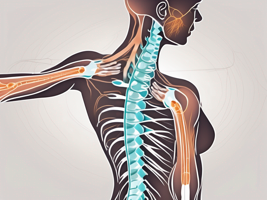
The accessory nerve, also known as cranial nerve XI, is a paired cranial nerve that plays a crucial role in controlling various head, neck, and shoulder movements. It originates from the medulla oblongata and spinal cord, consisting of two distinct components: the cranial part and the spinal part.
Anatomy of the Accessory Nerve
The cranial part of the accessory nerve emerges from the motor nucleus in the medulla, while the spinal part arises from the ventral horn of the upper cervical spinal cord segments. These two components join together to form the complete accessory nerve, which then courses through the posterior cranial fossa.
As the accessory nerve travels through the posterior cranial fossa, it passes through a small opening called the jugular foramen. This narrow pathway allows the nerve to enter the neck region, where it continues its journey to innervate specific muscles.
Descending through the neck, the accessory nerve provides motor innervation to two important muscles: the sternocleidomastoid and trapezius muscles. The sternocleidomastoid muscle, located on the sides of the neck, enables us to rotate our heads and tilt them sideways. On the other hand, the trapezius muscle, which covers a large portion of the upper back and neck, helps stabilize and move our shoulders.
It is fascinating to note the complex pathway that the accessory nerve takes to reach these muscles. This intricate network ensures that the nerve can influence a range of head, neck, and shoulder movements, allowing us to perform everyday tasks with ease.
Function of the Accessory Nerve
The primary function of the accessory nerve is to control the movement of the sternocleidomastoid and trapezius muscles. These muscles play significant roles in maintaining proper head and shoulder posture, as well as facilitating various movements.
Working in tandem, the sternocleidomastoid muscle allows us to rotate our heads and tilt them sideways, while the trapezius muscle helps stabilize and move our shoulders. These actions are essential for everyday activities such as looking around, reaching for objects, and maintaining balance.
However, the function of the accessory nerve goes beyond just controlling these two muscles. It also integrates with other cranial nerves and the cervical plexus, forming a complex network that ensures coordinated movements during various activities. This integration allows for the smooth execution of tasks that involve the neck and shoulders, such as turning our heads to follow a conversation, lifting heavy objects, or participating in physical activities.
In conclusion, the accessory nerve is a vital component of the nervous system, responsible for controlling the movement of the sternocleidomastoid and trapezius muscles. Its intricate pathway and integration with other nerves allow for the coordination of head, neck, and shoulder movements, enabling us to perform a wide range of activities with precision and ease.
The Roots of the Accessory Nerve
Defining Nerve Roots
Nerve roots, also known as rootlets, are the structural components that give rise to cranial and spinal nerves. They are essential for the proper functioning of the nervous system. In the case of the accessory nerve, it is formed by a combination of cranial and spinal roots.
The cranial root emerges directly from the medulla oblongata, which is located at the base of the brainstem. This root carries important motor fibers that are responsible for controlling the sternocleidomastoid muscle. This muscle plays a crucial role in various movements of the head and neck, such as rotating the head and flexing the neck.
On the other hand, the spinal roots of the accessory nerve originate from the upper cervical spinal cord segments. Specifically, they arise from the ventral horn, which is a region of the spinal cord that contains motor neurons. These spinal roots provide motor fibers that innervate the trapezius muscle. The trapezius muscle is a large muscle located in the upper back and neck region. It is responsible for movements such as shrugging the shoulders, pulling the shoulders back, and rotating the scapula.
It is fascinating to note that the cranial and spinal roots of the accessory nerve converge within the skull before exiting together through the jugular foramen. The jugular foramen is a small opening located at the base of the skull, near the junction of the temporal and occipital bones. This convergence of the roots allows for coordinated and synchronized control of the muscles they innervate.
Role of Nerve Roots in the Accessory Nerve
The cranial and spinal roots of the accessory nerve work in synergy to control the muscles they innervate. Each root has its own specific role and contributes to the overall function of the nerve.
The cranial root, originating from the medulla oblongata, provides motor fibers to the sternocleidomastoid muscle. This muscle is responsible for various movements of the head and neck, including turning the head from side to side and flexing the neck. The cranial root ensures that these movements are executed smoothly and precisely.
On the other hand, the spinal root of the accessory nerve supplies motor fibers to the trapezius muscle. The trapezius muscle is involved in a wide range of movements, such as lifting and rotating the shoulders, pulling the shoulders back, and maintaining proper posture. The spinal root ensures that the trapezius muscle functions effectively, allowing us to perform these movements with ease.
The coordination of these two roots highlights the complexity and versatility of the accessory nerve. By working together, they enable us to carry out a wide range of physical tasks that involve both the neck and the shoulders. Whether it’s turning our heads, shrugging our shoulders, or maintaining proper posture, the accessory nerve plays a crucial role in our everyday movements.
Understanding the roots of the accessory nerve provides us with valuable insights into the intricate workings of the nervous system. It is a testament to the remarkable complexity and precision of the human body, where even the smallest components contribute to our overall functionality.
Counting the Roots of the Accessory Nerve
Methodology for Identifying Nerve Roots
Determining the exact number of roots in the accessory nerve can be a challenging task. Researchers have employed various techniques to study the anatomy and root arrangement of this nerve.
Traditional methodologies include dissections, imaging techniques such as MRI and CT scans, and electrophysiological recordings. These approaches have provided insights into the structural complexity of the accessory nerve and its roots, allowing scientists and medical professionals to better understand its role in our motor function.
Dissections have been a valuable tool in exploring the intricate network of the accessory nerve. By carefully dissecting cadavers, researchers have been able to visualize the nerve roots and their connections. This hands-on approach provides a detailed understanding of the nerve’s structure and allows for precise identification of its roots.
Imaging techniques, such as MRI and CT scans, have revolutionized the field of neuroanatomy. These non-invasive methods allow researchers to visualize the accessory nerve and its roots in living individuals. By capturing high-resolution images, medical professionals can study the nerve’s morphology and identify any variations in root count.
Electrophysiological recordings have also played a crucial role in studying the accessory nerve. By measuring the electrical activity generated by the nerve, researchers can identify the different roots and assess their functionality. This technique provides valuable information about the nerve’s physiological properties and its contribution to motor control.
Number of Roots in the Accessory Nerve
While variability exists among individuals, the accessory nerve typically consists of one cranial root and one or more spinal roots. However, studies have reported cases where individuals possess additional cranial or spinal roots, indicating anatomical variations.
Understanding the variations in root count is essential for medical professionals. It allows them to anticipate potential challenges during surgical procedures or diagnose conditions that may affect the accessory nerve. By recognizing the possibility of additional roots, healthcare providers can tailor their treatment plans to meet the specific needs of each patient.
Furthermore, the presence of extra roots in the accessory nerve highlights the complexity of human anatomy. It serves as a reminder that our bodies are not always uniform and that individual variations can exist even in seemingly well-defined structures.
Therefore, it is crucial to recognize that the number of roots in the accessory nerve can vary from person to person. If specific medical or surgical interventions require precise knowledge of an individual’s accessory nerve root count, consulting with a medical professional becomes essential. They can utilize various diagnostic tools and techniques to accurately assess the root count and ensure the best possible outcome for the patient.
Implications of Accessory Nerve Root Count
Impact on Neurological Function
The number of roots in the accessory nerve may have implications for neurological function. Variations in root count can potentially affect the coordination and strength of the muscles innervated by the accessory nerve.
For example, individuals with a higher number of accessory nerve roots may have enhanced motor control and strength in the neck and shoulder region. This can contribute to improved performance in activities that require precise movements and stability, such as sports or playing a musical instrument.
On the other hand, individuals with a lower number of accessory nerve roots may experience challenges in coordinating movements and maintaining muscle strength in the affected areas. This can potentially lead to difficulties in performing certain tasks that require fine motor skills, such as writing or manipulating small objects.
Understanding an individual’s accessory nerve root count may be relevant in assessing certain conditions or injuries that involve the neck and shoulder region. For instance, individuals with a higher number of roots may have a lower risk of developing conditions such as thoracic outlet syndrome, which can cause pain, numbness, and weakness in the upper extremities.
Proper diagnosis and treatment planning can benefit from a comprehensive understanding of the accessory nerve’s anatomy and variation in root count. Healthcare professionals can tailor rehabilitation programs and interventions based on the individual’s specific root count, aiming to optimize their neurological function and overall quality of life.
Relevance for Surgical Procedures
Surgeons performing procedures in the neck and shoulder area must carefully consider the anatomy of the accessory nerve and its roots. Knowledge of root count can aid in surgical planning, minimizing the risk of unintended nerve damage during procedures such as neck dissections, tumor removals, and reconstructive surgeries.
During surgical procedures, the surgeon’s awareness of the accessory nerve root count can guide their approach to minimize potential complications. For instance, in cases where the accessory nerve has a higher number of roots, the surgeon may need to exercise greater caution to avoid injury to any additional branches.
Conversely, in cases where the accessory nerve has a lower number of roots, the surgeon may need to modify their surgical technique to ensure adequate preservation of the nerve’s function. This may involve adjusting the placement of incisions or using specialized instruments to minimize the risk of damage.
It is crucial that patients consult with their healthcare providers, particularly surgeons, to discuss the specific implications of accessory nerve root count and its potential impact on surgical outcomes. By having an open and informed discussion, patients can make well-informed decisions about their treatment options and have realistic expectations regarding their recovery and potential functional outcomes.
Common Misconceptions about the Accessory Nerve Roots
Debunking Myths about Nerve Root Count
There are various misconceptions surrounding the accessory nerve roots. One common misconception is that all individuals possess the same number of accessory nerve roots. However, as discussed earlier, anatomical variations exist, and the number of roots can differ from person to person.
It is essential to dispel these myths to prevent misconceptions from influencing medical decisions or causing unnecessary concern. Consulting with medical professionals and relying on scientific evidence can help clarify any misunderstandings about accessory nerve root count.
Furthermore, the number of accessory nerve roots can vary not only between individuals but also within the same person. Studies have shown that some individuals may have asymmetrical accessory nerve root counts, with one side of the body having more roots than the other. This variability highlights the complexity of the human nervous system and emphasizes the importance of considering individual differences when assessing nerve function.
Moreover, the number of accessory nerve roots is not the sole determinant of nerve function. While it is true that the accessory nerve plays a crucial role in motor control, its function is not solely dependent on the number of roots. Other factors, such as the integrity of the nerve fibers and their connections to the muscles, also contribute to the overall functioning of the accessory nerve.
Clarifying Confusions about Accessory Nerve Structure
Another misconception relates to the exact organization and arrangement of the accessory nerve roots. The complex pathway of the accessory nerve, combined with its variable root count, can lead to confusion about its structure and function.
Medical professionals play a crucial role in clarifying these confusions by providing accurate information and addressing any concerns or questions related to the anatomy and function of the accessory nerve roots.
Furthermore, understanding the accessory nerve’s structure requires a comprehensive knowledge of its course through the body. The accessory nerve roots originate from the spinal cord’s upper segments, specifically the cervical spinal nerves. From there, they ascend through the foramen magnum, a large opening at the base of the skull, before entering the cranial cavity.
Once inside the cranial cavity, the accessory nerve roots join with the cranial nerve XI, forming a complex network of nerve fibers. This intricate network then extends downward, passing through the jugular foramen, a narrow opening in the skull’s base.
It is important to note that the accessory nerve roots do not function in isolation. They work in conjunction with other cranial nerves and spinal nerves to control various muscles involved in head and neck movements. This collaborative effort ensures the smooth execution of motor functions and highlights the interconnectedness of the nervous system.
Future Research Directions on Accessory Nerve Roots
Unanswered Questions about the Accessory Nerve
Despite ongoing research and advancements in neuroscience, several questions about the accessory nerve roots and their significance remain unanswered.
Future research endeavors aim to explore aspects such as the specific roles of individual roots, the impact of anatomical variations on motor function, and potential correlations between root count and pathological conditions affecting the accessory nerve.
Potential Areas for Further Investigation
Areas for further investigation include the development of non-invasive techniques to accurately determine accessory nerve root count and the influence of accessory nerve variations on physical therapy outcomes for patients with neck and shoulder-related conditions.
With continued research and collaboration among scientists, medical professionals, and allied healthcare experts, our understanding of the accessory nerve roots will undoubtedly deepen, leading to enhanced clinical management and improved patient outcomes.
In conclusion, the accessory nerve, with its intricate root arrangement, plays a vital role in our motor functions. While most individuals possess one cranial and one or more spinal roots, variations in root count are possible. Understanding the implications of accessory nerve root count is crucial in medical assessments and surgical planning. By debunking misconceptions and focusing on future research, we can expand our knowledge of accessory nerve roots, ultimately benefiting patients and advancing medical science.


