accessory nerve comes form which cervical nerves

The accessory nerve is a crucial component of the human nervous system, playing a significant role in motor function. In order to understand the accessory nerve and its functions fully, it is important to delve into its anatomy, explore its connection with the cervical nerves, and examine its clinical significance, treatment, and management.
Understanding the Accessory Nerve
The accessory nerve, also known as the spinal accessory nerve or cranial nerve XI, is one of the 12 pairs of cranial nerves. It originates from the medulla oblongata, the lower part of the brainstem, and extends downwards into the spinal cord.
The accessory nerve plays a crucial role in the functioning of the human body. Let’s dive deeper into its anatomy and function to gain a comprehensive understanding.
Anatomy of the Accessory Nerve
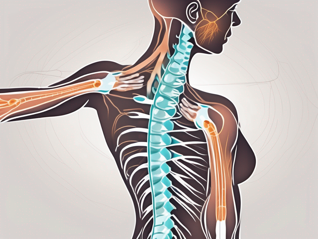
Comprised of both cranial and spinal components, the accessory nerve has a complex anatomical structure. The cranial portion arises from the nucleus ambiguous within the medulla oblongata, while the spinal portion emerges from the upper five or six cervical spinal nerves.
The cranial component of the accessory nerve travels through the skull and joins the vagus nerve, another cranial nerve, to form a loop known as the jugular foramen. This intricate connection allows for coordinated motor control of various muscles involved in speech, swallowing, and head movements.
On the other hand, the spinal component of the accessory nerve descends into the neck, passing through the posterior triangle. It innervates the sternocleidomastoid and trapezius muscles, which are essential for proper neck and shoulder movements.
The accessory nerve’s unique dual origin and distribution make it a vital player in the complex network of nerves responsible for motor control in the head, neck, and upper body.
Function of the Accessory Nerve
The primary function of the accessory nerve is to control the movement of certain muscles, including the sternocleidomastoid and trapezius muscles. These muscles are vital for actions such as head rotation, shoulder elevation, and upper back control.
When the accessory nerve is functioning properly, it allows for smooth coordination and precise control of these muscles. This enables us to perform a wide range of activities, from turning our heads to lifting heavy objects.
However, certain conditions or injuries can affect the accessory nerve, leading to motor impairments. Damage to the cranial component of the accessory nerve may result in difficulties with speech and swallowing, while damage to the spinal component can cause weakness or paralysis in the neck and shoulder muscles.
Rehabilitation techniques, such as physical therapy and targeted exercises, can help individuals recover and regain optimal functioning of the accessory nerve. Understanding the intricate role this nerve plays in our daily movements is crucial for healthcare professionals and patients alike.
In conclusion, the accessory nerve is a remarkable cranial nerve that facilitates essential motor control in the head, neck, and upper body. Its complex anatomy and function make it a fascinating subject of study for anatomists, neurologists, and anyone interested in the intricacies of the human nervous system.
The Cervical Nerves: An Overview
In order to understand the origin of the accessory nerve, it is essential to have a basic understanding of the cervical nerves.
The cervical nerves, also known as the cervical spinal nerves, emerge from the spinal cord in the neck region. They are responsible for transmitting sensory and motor information between the brain and various parts of the body, including the neck, shoulders, and arms.
These nerves play a crucial role in the functioning of the nervous system. They serve as the communication pathway between the brain and the rest of the body, allowing for the transmission of signals that control movement, sensation, and other bodily functions.
Each cervical nerve is unique in its function and distribution. There are eight pairs of cervical nerves labeled C1 to C8. These nerves innervate specific areas of the body and have distinct functions.
The Role of Cervical Nerves in the Nervous System
The cervical nerves are integral components of the nervous system. They serve as the connection between the brain and the peripheral nervous system, allowing for the transmission of signals to and from various parts of the body.
These nerves are responsible for transmitting sensory information from the neck, shoulders, and arms to the brain. This includes sensations such as touch, temperature, and pain. They also play a crucial role in motor function, allowing for the control of movement in these areas.
Furthermore, the cervical nerves are involved in the autonomic nervous system, which regulates involuntary bodily functions such as heart rate, blood pressure, and digestion. They help maintain homeostasis by coordinating the body’s response to internal and external stimuli.
Differentiating Between Cervical Nerves
Each cervical nerve has its own distinct set of functions and distributions. Understanding these differences is essential in diagnosing and treating conditions that affect specific areas of the body.
The first cervical nerve, C1, is responsible for innervating the back of the head and the neck. It plays a crucial role in maintaining proper posture and head movement.
C2 and C3 are primarily involved in sensory innervation of the scalp, face, and neck. They transmit information related to touch, temperature, and pain from these areas to the brain.
C4 is responsible for innervating the shoulder region, including the upper part of the arms. It plays a role in controlling movements such as lifting and rotating the shoulders.
C5, C6, C7, and C8 are involved in innervating the upper limbs, including the arms and hands. They transmit sensory information and control motor function in these areas.
Understanding the functions and distributions of the cervical nerves is crucial in diagnosing and treating conditions such as cervical radiculopathy, which is characterized by pain, weakness, and numbness in specific areas of the neck and upper limbs.
In conclusion, the cervical nerves are essential components of the nervous system. They play a crucial role in transmitting sensory and motor information between the brain and various parts of the body. Each cervical nerve has its own unique set of functions and distributions, allowing for precise control and coordination of movement and sensation. Understanding the intricacies of these nerves is vital in diagnosing and treating conditions that affect specific areas of the body.
The Origin of the Accessory Nerve
The accessory nerve, despite its unique functions, has an intriguing connection with the cervical nerves from which it originates.
The accessory nerve, also known as cranial nerve XI, is a motor nerve that plays a crucial role in controlling certain muscles in the head and neck. It is responsible for movements such as turning the head, shrugging the shoulders, and lifting the arms. While it has its own distinct functions, its origin lies in the cervical nerves.
The Connection Between Accessory Nerve and Cervical Nerves
The accessory nerve’s spinal component emerges from the upper five or six cervical nerves, specifically C1 to C6. This intimate connection highlights the interplay between the accessory nerve and the cervical nerves in motor control and coordination.
Within the spinal cord, the fibers of the accessory nerve merge with the fibers of the cervical nerves, forming a complex network that allows for the transmission of signals between the brain and the muscles of the head and neck. This intricate connection ensures the smooth and coordinated movement of these muscles, enabling us to perform various actions with precision.
Interestingly, the accessory nerve is unique among the cranial nerves as it has both cranial and spinal components. While the cranial portion originates in the brainstem, the spinal portion arises from the cervical nerves. This dual origin further emphasizes the close relationship between the accessory nerve and the cervical nerves.
How the Accessory Nerve Develops from Cervical Nerves
During embryonic development, the accessory nerve and cervical nerves share common developmental pathways. This intermingling explains why damage to the cervical nerves can occasionally lead to dysfunction of the accessory nerve.
As the embryo develops, the neural crest cells give rise to both the accessory nerve and the cervical nerves. These cells migrate and differentiate into the specialized nerve cells that form the intricate network of the nervous system. The close proximity of the developing accessory nerve and cervical nerves allows for their mutual influence on each other’s growth and development.
Any disruption or injury to the cervical nerves during this critical developmental period can potentially affect the proper formation and function of the accessory nerve. This can result in various motor deficits and coordination issues, highlighting the delicate nature of the accessory nerve’s origin.
In conclusion, the accessory nerve’s origin in the cervical nerves showcases the intricate relationship between these structures in motor control and coordination. Understanding the developmental and anatomical connections between the accessory nerve and cervical nerves provides valuable insights into the functioning of the head and neck muscles and the potential consequences of any disruptions in this complex network.
Clinical Significance of the Accessory Nerve
Understanding the clinical significance of the accessory nerve is crucial in order to identify potential disorders or damage that may occur.
The accessory nerve, also known as the eleventh cranial nerve, plays a vital role in the functioning of various muscles in the head, neck, and shoulders. It is responsible for innervating the sternocleidomastoid and trapezius muscles, which are essential for movements such as head rotation, shoulder shrugging, and maintaining proper posture.
Disorders Related to the Accessory Nerve:
Damage or dysfunction of the accessory nerve can result in various disorders, such as accessory nerve palsy. This condition can lead to weakness or paralysis of the affected muscles, making it difficult for individuals to perform everyday activities that require head or shoulder movements. For example, turning the head to check blind spots while driving or lifting objects overhead may become challenging.
In addition to accessory nerve palsy, other conditions may also affect the accessory nerve. Nerve impingement or entrapment can occur due to compression or pressure on the nerve, leading to discomfort and limited mobility. This can cause symptoms such as pain, tingling, or numbness in the neck, shoulders, or upper back.
Diagnostic Techniques for Accessory Nerve Damage:
Medical professionals employ different diagnostic techniques to assess accessory nerve disorders. These techniques aim to accurately identify the underlying cause of the symptoms and determine the extent of nerve damage. A thorough physical examination is often the first step, where the healthcare provider evaluates muscle strength, range of motion, and any signs of muscle wasting or atrophy.
In addition to the physical examination, electromyography (EMG) may be used to assess the electrical activity of the muscles innervated by the accessory nerve. This test involves the insertion of small electrodes into the muscles to measure their response to nerve signals. Abnormalities in the EMG results can indicate nerve damage or dysfunction.
Imaging studies such as magnetic resonance imaging (MRI) may also be utilized to visualize the accessory nerve and surrounding structures. MRI can provide detailed images of the nerves, muscles, and other soft tissues, helping to identify any structural abnormalities or potential causes of nerve compression.
In conclusion, understanding the clinical significance of the accessory nerve is crucial for healthcare professionals to diagnose and manage disorders related to this important cranial nerve. By utilizing various diagnostic techniques, they can accurately assess the extent of nerve damage and develop appropriate treatment plans to improve the quality of life for individuals affected by accessory nerve disorders.
Treatment and Management of Accessory Nerve Disorders
Once an accessory nerve disorder is diagnosed, appropriate treatment and management strategies can be implemented to improve the patient’s quality of life.
Accessory nerve disorders can significantly impact a person’s daily functioning and overall well-being. Therefore, it is crucial to explore various treatment options to address the specific needs of each individual.
Surgical Interventions for Accessory Nerve Disorders
In severe cases, surgical intervention may be necessary to repair or reconstruct the damaged accessory nerve. This procedure is typically performed by a skilled neurosurgeon and requires careful consideration of the patient’s specific condition.
The decision to undergo surgery for an accessory nerve disorder is not taken lightly. It involves a thorough evaluation of the risks and benefits associated with the procedure. The neurosurgeon will assess the extent of nerve damage, the potential for nerve regeneration, and the overall health of the patient before recommending surgery.
During the surgical intervention, the neurosurgeon may use various techniques to repair or reconstruct the damaged accessory nerve. These techniques may involve nerve grafts, nerve transfers, or other innovative approaches to restore proper nerve function.
Following the surgery, patients will typically undergo a period of recovery and rehabilitation. Physical therapy and other supportive measures may be recommended to optimize the outcomes of the surgical intervention.
Rehabilitation and Physical Therapy for Accessory Nerve Damage
Physical therapy and rehabilitation programs can be tremendously beneficial in restoring functionality and improving strength in individuals with accessory nerve damage. These programs often involve a combination of exercises, stretches, and other therapeutic techniques tailored to each patient’s unique needs.
The goal of rehabilitation and physical therapy is to enhance the patient’s range of motion, promote muscle strength and coordination, and improve overall functional abilities. This may involve exercises targeting specific muscle groups affected by the accessory nerve disorder.
Physical therapists and rehabilitation specialists work closely with patients to develop personalized treatment plans that address their specific impairments and goals. These plans may include exercises to improve shoulder and neck mobility, strengthen weakened muscles, and enhance overall motor control.
In addition to physical therapy, other complementary therapies may be incorporated into the treatment plan. These may include occupational therapy, massage therapy, acupuncture, or electrical stimulation techniques to further promote nerve regeneration and recovery.
While it is informative to understand the accessory nerve’s connection to the cervical nerves, it is essential to avoid self-diagnosis or self-treatment. If you suspect any issues related to the accessory nerve, it is crucial to consult with a medical professional for an accurate diagnosis and appropriate treatment plan. An experienced healthcare provider will guide you through the necessary steps to ensure optimal management of any accessory nerve disorders and improve your overall well-being.


